Poster
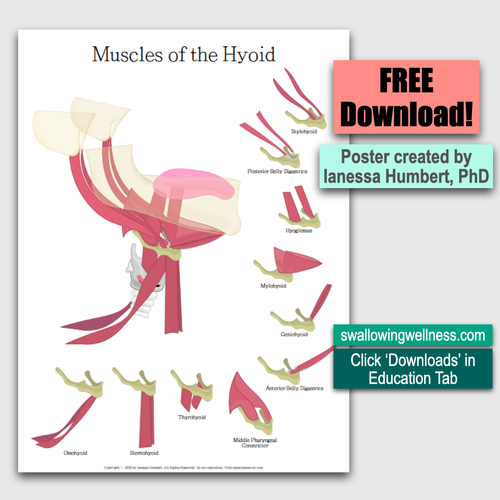
Muscles of the Hyoid Bone
Learn all the muscles of the hyoid bone through Dr. Humbert’s original poster.
Download the original PDFTeaching library
A local home for Dr. Humbert's visual teaching materials and concise clinical topics that previously lived on the retiring site.
Current format
Three original PDFs available for direct download
01
These are the exact PDFs previously delivered only after submitting a form on the source site. They now download directly, without collecting a name or email address.
Poster
Learn all the muscles of the hyoid bone through Dr. Humbert’s original poster.
Download the original PDF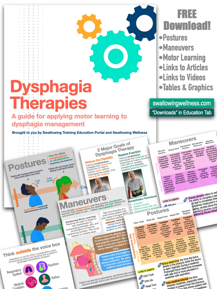
Clinical handout
The original handout on traditional postures and maneuvers, compensation, rehabilitation, motor learning, and neuroplasticity.
Download the original PDF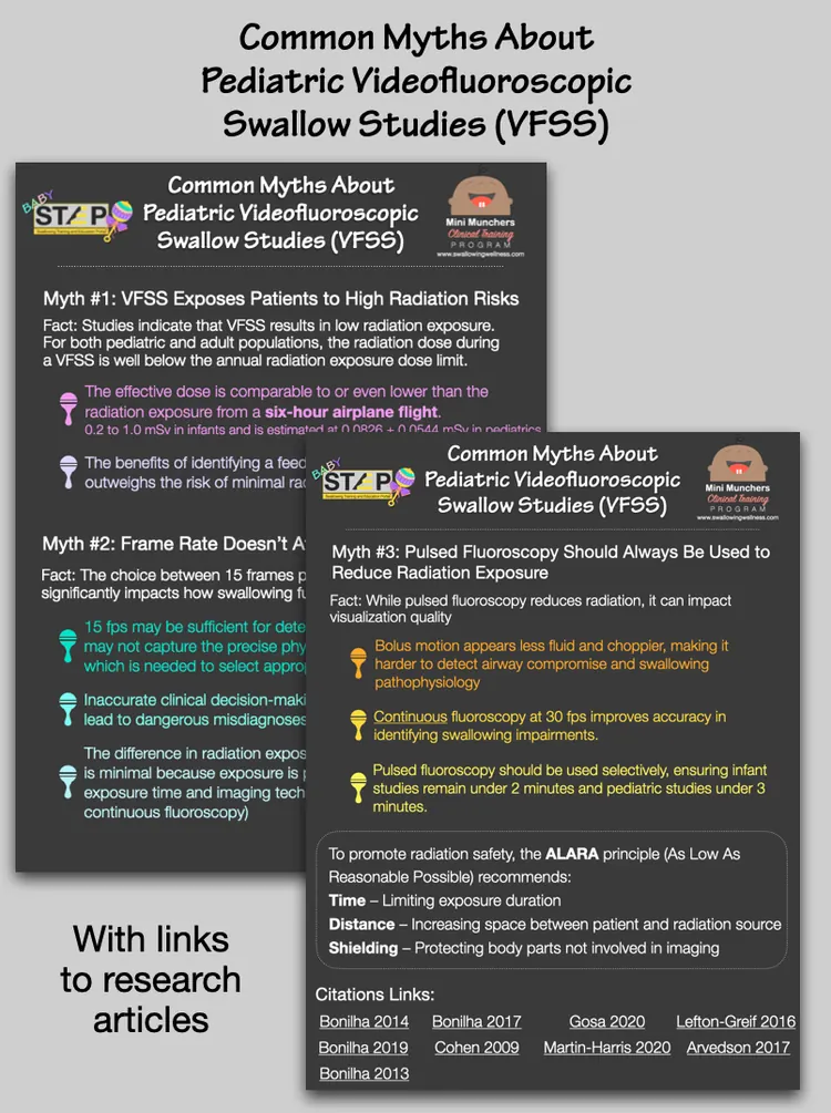
Two-page handout
The original evidence-focused handout on radiation risk, frame rate, and pulsed fluoroscopy in pediatric videofluoroscopic swallow studies.
Download the original PDF02
Since we have made the hyoid bone the celebrity of swallowing physiology, we should at least understand it. The hyoid bone is not a fan of other bones: it is the only bone in the human body that does not directly articulate with or connect to another bone. It is also known as the “tongue bone.”
It has more connections to a wider variety of external structures than any other bone, including the larynx, jaw, tongue, pharynx, skull, sternum, and scapula (shoulder blade). Given all of its connections to surrounding structures, it lives in a muscular throne.
Think of it as a royal puppet. Its movement indicates which strings—muscles—are influencing its direction and position in the neck. For example, if it moves toward the jaw, then the suprahyoid, submental, and floor-of-mouth muscles (mylohyoid, geniohyoid, and anterior belly of the digastrics) are contracting. If it moves toward the sternum, then the infrahyoid muscles (sternohyoid and omohyoid) are moving it.
It moves during speech, swallowing, coughing, yawning, and almost all behaviors within the head and/or neck.
During swallowing it follows an upward-forward, boomerang-like arc. The extent of this excursion is quite variable in normal swallowing (Molfenter et al., 2011). Hyoid forward movement ranges from 8 to 18 mm, and hyoid upward movement ranges from 6 to 25 mm.
Clinical relevance: it is an important muscular attachment for many structures in the head and neck and is therefore involved in many head-and-neck functions. Expert understanding of swallowing physiology involves extrapolating hyoid movement to infer the muscle groups influencing its direction.
A rudimentary and misguided interpretation labels hyoid movement normal or abnormal without understanding why it moved, what normal is, or whether the movement affected the swallow—and uses palpation alone to guess at adequate hyoid function. Studies show that clinicians may identify “abnormal” hyoid movement when the hyoid is moving within normal ranges (Brates et al., 2019).
The study cited in the original text about clinical judgments of hyoid movement.
Open PMID 3004308003
The Dysphagia Therapy handout explains traditional postures and maneuvers used as either compensatory strategies or rehabilitative techniques. Motor learning and neuroplasticity principles are highlighted to differentiate between treatments that do not challenge the patient—reducing disability—versus those that challenge the swallowing impairment with the goal of restoring swallowing function.
Links to relevant research literature and video examples are included. The handout is sponsored by STEP Community and Swallowing Wellness.
The source handout page directs clinicians interested in in-person, hands-on therapy training to the Swallow Treat Retreat.
Explore the Treat RetreatThe source page also directs clinicians to STEP for high-quality, low-cost online training.
Visit STEP04
Misunderstandings about pediatric videofluoroscopic swallow studies (VFSS), also known as modified barium swallow studies (MBSS), can lead to poor clinical decision-making that may be dangerous for infants and children with pediatric feeding and swallowing disorders.
05
Open each image at full size for teaching, discussion, or closer review of the citations included in the artwork.
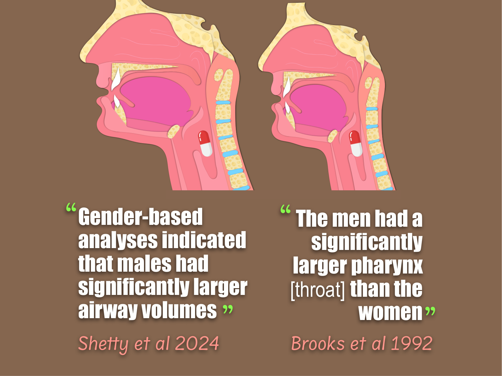
Research in practice
A research teaching graphic comparing published findings about anatomy by sex, with cited studies shown in the artwork.
Open full-size graphic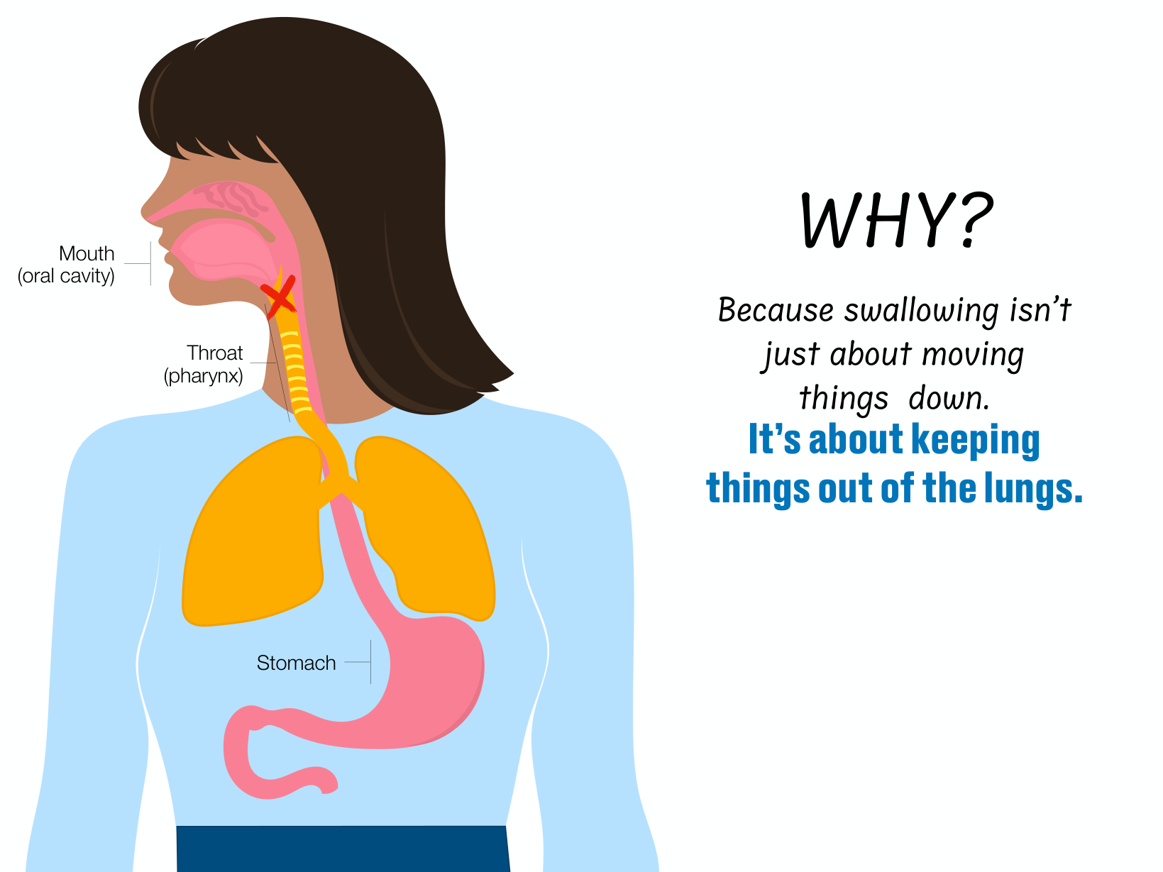
Airway protection
A labeled teaching graphic for discussing the airway, food pathway, and the structures involved in airway protection.
Open full-size graphic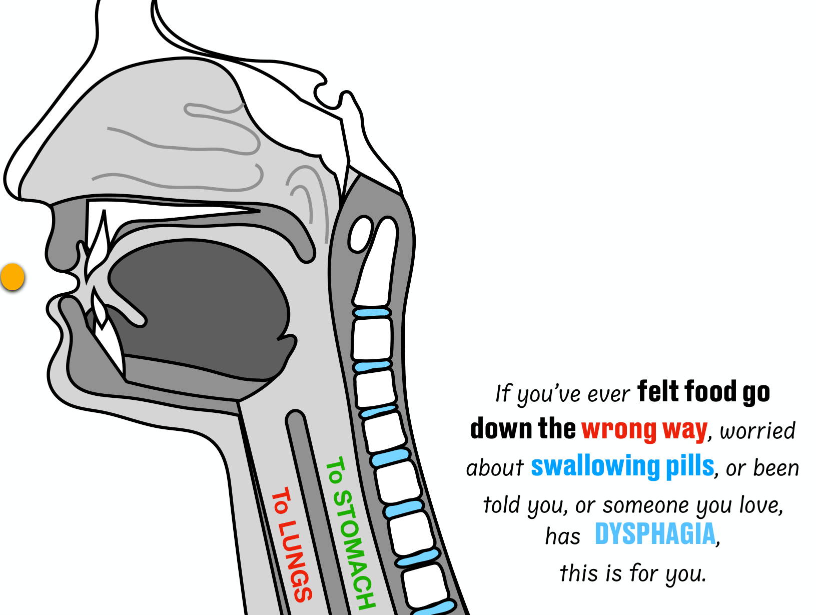
Anatomy
A visual reference for orienting learners to the neighboring breathing and swallowing pathways.
Open full-size graphic